

Files
Download Full Text (423 KB)
Program
Emergency Medicine
Training Level
Resident PGY 3
Institution
Henry Ford Macomb
Abstract
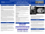
Background: Portal venous thrombosis (PVT) can be generally classified into three categories- acute non cirrhotic PVT, chronic PVT (also called extrahepatic portal venous occlusion), and PVT in cirrhosis. PVT is usually seen in those with liver disease and it is rare in patients without. Management is usually with anticoagulation and finding and treating the etiology of the PVT. If untreated, patients can develop portal hypertension. This patient presented a unique diagnostic challenge as she had risk factors for numerous etiologies of PVT. Case description: A 64 year old female with a history of hypertension, hyperlipidemia, and a lung nodule presented to the emergency department with fatigue, fever, nausea, and vomiting. She sought medical care upon returning home from a cruise in Mexico. She was febrile and tachycardic and, though she did not have any abdominal tenderness, stated that a few days prior she did have right upper quadrant abdominal pain. CT scan with IV contrast was ordered and showed a masslike area in the anterior left hepatic lobe, acute left PVT, and enlarged porta hepatis lymph nodes. Subsequently, an MRCP was ordered as there was concern for malignancy given the CT findings which redemonstrated the left portal venous thrombosis and the mass described on the CT was further determined to be hepatic periportal edema. Shortly after presentation she was started on broad spectrum antibiotics given concern for sepsis as well as anticoagulation for the PVT. Blood cultures grew streptococcus intermedius, strepconstellatus, and eikenella corrodens. Given a dental procedure two months prior, a TEE was performed which did not show vegetation and endocarditis was ruled out. Given a family history of colon cancer she had a colonoscopy which showed diverticulosis with friable mucosa. That was ultimately thought to the be etiology of her bacteremia. Discussion: This case was interesting in that the patient had risk factors for multiple etiologies of her sepsis and PVT. She had recent dental work, recent travel on the cruise, a history of a lung nodule, family history of colon cancer, initial CT scan with concern for liver mass and possible metastatic disease. She had a thorough work up that ultimately led to the thought that her bacteremia was due to diverticular disease and her sepsis was likely the etiology of her PVT. She was placed on antibiotics for 4 weeks outpatient as well as started on oral anticoagulation with a plan for repeat imaging of the PVT at the three month mark to determine length of anticoagulation.
Presentation Date
5-2019
Recommended Citation
Saghy, Nicole and Davis-Baumann, Jillian, "Acute portal venous thrombosis caused by severe sepsis and not associated with liver disease" (2019). Case Reports. 106.
https://scholarlycommons.henryford.com/merf2019caserpt/106
