

Files
Download Full Text (9.0 MB)
Program
Internal Medicine
Training Level
Resident PGY 2
Institution
Henry Ford Hospital
Abstract
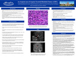
Introduction: Atypical Teratoid Rhabdoid tumors are rare pediatric tumors that usually occur at age <3 years. These tumors are scarcely seen in adults, with the first adult case appearing in 1992. Since then, about 64 cases have been reported in the literature. As such, much what has been learned about adultATRT cases has come from case reports and extrapolation frompediatric cases. The loss of INI1 (SMARCB1) or BRG1 (SMARCA4) genes are implicated in pathogenesis of ATRT3. Specifically, the INI1/SMARCB1 gene is classified as a tumor-suppressor gene that encodes a core subunit protein of the ATP-dependent SWI/SNF chromatin remodeling complex. Case Presentation: A 62-year-old Caucasian right-handed female with history of hypertension and sinusitis presented with 2-month history of bilateral, frontal headaches reaching 5/10 in severity with associated nausea, emesis, polydipsia and polyuria. Polyuria occurred every hour while polydipsia included drinking (15-20) 16 oz. bottles per day.She presented to a local hospital and was found to have hypernatremia (Sodium 154 mEq/mL). Non-contrast brain computerized tomorgraphy (CT) revealed a 1.2cm x 1.1cm x 1.7 cm sellar mass with suprasellar extension. Urine studies diagnosed central diabetes insipidus, responsive toD-amino D-arginine vasopressin (DDAVP). However, she developed seizures and Abducen’s nerve palsy. Magnetic resonance imaging (MRI) of the brain demonstrated intraventricular and subarachnoid hemorrhage along with optic nerve edema. Prior to surgery,she was found on the floor of her room in pool of urine with incoherent speech and a sluggish pupillary reflex on right side. STAT non-contrast head CT revealed 2.7 x 1.8 x 2.5 cm extension of hemorrhage into interpeduncular cisterns and ventricles with associated 3rd ventricle & lateral ventricle. There was no midline shift or impending herniation. She received bi-coronal craniotomy with excision of sellar mass and right frontal external ventricular drain placement. Hydrocephalus and hemorrhage improved on subsequentMRI a few days later. The pathology report came back positive for a malignant epithelioid neoplasm, specifically sellar ATRT, WHO grade V. Tumor was shown to be SMARCB1/INI1 deficient and no metastatic lesions were found. Recommendations were made for craniospinal radiation and chemotherapy afterward. Discussion: ATRTs remain rare and aggressive brain tumors seen in both the pediatric and adult population. The management of ATRT remains a difficult challenge with multimodal approaches to treatment remaining the mainstay. Resection followed by radiation and chemotherapy has been shown to significantly increase 5-year overall survival rate, yet median time to progression remains in the range of 6-10 months. Much more standardization is required in the treatment of disease, since patients continue to get variable approaches to treatment. Additionally, radiation doses and optimal chemotherapy regimens have yet to be determined. A promising step towards these answers have been in-vitro studies of Insulin-growth factor receptor (IGF-1R) inhibition in sensitizingthe tumor to chemotherapy and radiation.
Presentation Date
5-2019
Recommended Citation
Mahmood, Sharmeen and Mohammed, Hadi, "An Atypical Case of Atypical Teratoid Rhabdoid Tumor (ATRT)" (2019). Case Reports. 43.
https://scholarlycommons.henryford.com/merf2019caserpt/43
