

Files
Download Full Text (266 KB)
Program
Radiology - Diagnostic
Training Level
Resident PGY 2
Institution
Henry Ford Hospital
Abstract
Background: Immune checkpoint inhibitors have emerged as a promising new class of anti-cancer drugs. Unfortunately, while potentiating the immune system’s response to cancer cells, these drugs also place the patient at risk for the development of immune-related adverse events. Although most commonly involving the gastrointestinal tract and skin, involvement of the endocrine system also occurs, with hypophysitis one of the most frequent complications. This case report describes a case of immune checkpoint inhibitor-induced hypophysitis in a patient with metastatic melanoma undergoing combination therapy, and the associated imaging findings.
Case Report: A 60-year-old male undergoing combination immune checkpointtherapy with nivolumab (an anti-PD-1 antibody) and ipilimumab (an anti-CTLA-4 antibody)formetastatic melanoma developed headaches. An MRI of the brain was obtained and demonstrated new enlargement of the pituitary. The patient was also found to be hypothyroid. Further endocrinological evaluation revealed low testosterone, low random cortisol level, and low TSH, consistent with pituitary insufficiency. At this time, the checkpoint inhibitor therapy was held and the patient was treated with prednisone with excellent clinical response, including resolved headaches and improved energy. Follow-up MRI demonstrated resolution of the pituitary enlargement.
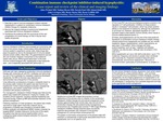
Imaging Findings: Sagittal pre-contrast MR images demonstrate a normal appearance of the pituitary prior to initiation of the combination checkpoint inhibitor therapy and interval enlargement of the pituitary approximately three months later near the end of the combination therapy. A sagittal post-contrast MR image following checkpoint inhibitor therapy discontinuation and steroid treatment demonstrates near resolution of the pituitary enlargement. Conclusion: As new cancer therapies emerge, physicians must remain aware of the potential for unique adverse effects – such as those occurring with immune checkpoint inhibitor therapies, including hypophysitis. In addition, as the imaging appearance of hypophysitis is not specific, understanding the clinical context in which it occurs can aid radiologists in recognizing it as a possible diagnosis and help guide appropriate clinical management.
Presentation Date
5-2019
Recommended Citation
Hyson, Nathan; Wrubel, Allen; Patel, Suresh C.; Dalal, Ishani; Corrigan, John; Marin, Horia; and Griffith, Brent, "Combination immune checkpoint inhibitor-induced hypophysitis: A case report" (2019). Case Reports. 83.
https://scholarlycommons.henryford.com/merf2019caserpt/83
