

Files
Download Full Text (444 KB)
Program
Internal Medicine
Training Level
Resident PGY 2
Institution
Henry Ford Hospital
Abstract
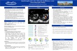
Introduction: Quadriscupidaortic valve (QAV) is a rare congenital heart defect typically found incidentally without any associated cardiac defects. The functional status of QAV is pure aortic insufficiency (AI), however, clinical manifestations are dependent on the functional status of the valve, presenting in the fifth or sixth decade of life due to progressive degeneration of the leaflets. In our case, we present a 37-year-old female who developed post-partum dyspnea with elevated brain natriuretic peptide (BNP) levels concerning for heart failure. Transesophageal echocardiography (TEE) revealed a preserved ejection fraction with aortic regurgitation consistent with valvular heart failure, however, incidentally showed a QAV.
Case Presentation: A 37-year-old female 5 days post cesarean section presented with dyspnea on exertion. Her physical examination was significant for a decrescendo diastolic murmur at the aortic area and bibasilar rales. Pertinent labs revealed a BNP level elevation of 345 pg/mL with normal troponin levels. Given her symptoms and elevated biomarkers, a transthoracic echocardiogram was obtained and was suggestive of AI. She was referred for transesophageal echocardiogram for better visualization of the aortic valve. TEE revealed a QAV with all four leaflets equal in size with normal thickness and mobility. Moderate malcoaptation of all valves was present and severe AI was visualized. Planimetry of aortic regurgitant orifice was measured at 0.29 cm2, the AI jet was greater than 65% of left ventricular outflow tract, pressure half time calculated at 252 ms, and venacontracta measured 0.6 cm. Her systolic (ejection fraction 60%) and diastolic function were both preserved. After diuresis, she was discharged home and followed up with structural heart and cardiac surgery. She was to have a CT coronary angiography performed part of her pre-operative evaluation, but was lost to follow up.
Discussion: QAV is a rare congenital cardiac anomaly that is typically found incidentally. The most prevalent complication of QAV is AI, however, these patients are also at increased risk for infective endocarditis. This is due to the progressive degeneration of the leaflets from the asymmetric mechanical stress around the four cusps. Echocardiography allowed for visualization of the aortic valve and for quantification of the degree of AI. Given her clinical presentation and cardiac risk factors, it was unclear what was causing her symptoms on admission. Through echocardiography, a diagnosis was made and the patient was able to receive appropriate care. The advancement in imaging techniques has increased the capability to diagnose QAV and its complications. The definitive treatment of QAV with AI is valve replacement, which was recommended to our patient.
Conclusion: QAV is a rare congenital disease that most commonly manifests with AI. QAV is typically found incidentally in the fifth and sixth decade of life and best visualized by TEE. Definitive management of QAV is valve replacement.
Presentation Date
5-2020
Recommended Citation
Demertzis, Zachary; Nona, Paul; and Zweig, Bryan, "Quadricuspid aortic valve with aortic insufficiency: a rare echocardiographic finding" (2020). Case Reports. 100.
https://scholarlycommons.henryford.com/merf2020caserpt/100
