

Files
Download Full Text (271 KB)
Program
Ophthalmology
Training Level
Resident PGY 3
Institution
Henry Ford Hospital
Abstract
Introduction: Thiamine is an important vitamin that serves as a crucial cofactor for glucose metabolism. As a result, thiamine deficiency affects many organ systems and can lead to significant morbidity and mortality. The brain heavily relies on glucose metabolism and is thus particularly susceptible to thiamine deficiency. Consequently, thiamine deficiency can lead to Wernicke’s encephalopathy (WE), a clinical syndrome consisting of the triad of ataxia, confusion, and ophthalmic abnormalities (Wei-Yi et al. 2013). At times, ophthalmologic manifestations, which include optic neuropathy, ophthalmoparesis, and nystagmus, provide the first clue of a thiamine deficiency. Optic neuropathy is the rarest presentation, only present in about 2.6% of patient with WE (Wei-Yi et al. 2013). Often times, the ophthalmic complications of thiamine deficiency present to the clinician concurrently. We present a case report that highlights the unique rapid progression of these ophthalmic manifestations that instead occurs over the course of several days.
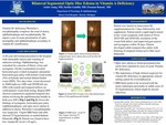
Case Report: A 48 year old woman initially presented to the hospital with intractable nausea and vomiting of unknown etiology. The patient also developed blurry vision during her hospitalization. Ocular examination revealed horizontal gaze evoked nystagmus and partial horizontal gaze palsy. Near vision was JI in both eyes (OU) at bedside, with intact fields to confrontation and normal dilated fundus examination (DFE). Her symptom of blurriness was initially attributed to her nystagmus. Two days later, the vision decreased to 20/60 right eye (OD) and 20/200 left eye (OS). The patient’s gaze palsy had worsened and she now had new central and bitemporal field defects on confrontation. A formal visual field test could not be done due to severe, persistent vomiting and mild encephalopathy. Repeat DFE showed symmetrical segmental temporal disc edema involving the papillomacular bundle OU (Figure 1). This was confirmed on optical coherence tomography (OCT) imaging (Figure 2). Given the triad of nystagmus, horizontal gaze palsy, and optic nerve edema, magnetic resonance imaging (MRI) of the brain and orbit was urgently done which showed T2 hyperintensity of the medial thalami bilaterally. The thiamine level was reduced to <10 >μg/L. A presumptive diagnosis of WE was made and patient was started on intravenous thiamine for three days followed by oral supplementation. Patient noticed a rapid improvement in her visual symptoms, with return of VA to 20/25 OD and 20/20 OS, resolution of the visual field defect, and improved gaze paresis and nystagmus. There was resolution of her optic nerve edema OU (Figures 1 and 2). During her subsequent outpatient follow ups, she developed subtle temporal disc pallor with thinning of her temporal retinal nerve fiber layers bilaterally.
Conclusion: The findings from this case report emphasize the importance of having a high clinical suspicion for thiamine deficiency in the setting of initial non-specific symptoms. It is vital to consider thiamine deficiency even in a patient without the usual risk factors such as alcoholism or gastric bypass surgery (Gratton and Lam 2014). This case also potentially demonstrates that ocular motor signs and symptoms can be a precursor to the more serious optic neuropathy that can present from thiamine deficiency. By understanding the possible step-wise progression of ophthalmic abnormalities as highlighted in this case report, clinicians may be able to prevent clinically significant visual loss.
Presentation Date
5-2020
Recommended Citation
Aung, Andre; Gandhi, Sachin; and Bansal, Poonam, "Bilateral Segmental Optic Disc Edema in Vitamin B1 Deficiency" (2020). Case Reports. 112.
https://scholarlycommons.henryford.com/merf2020caserpt/112
