

Files
Download Full Text (204 KB)
Program
Wayne State University Medical School
Training Level
Medical Student
Institution
Wayne State University
Abstract
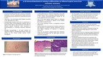
In the monitoring of patients who have had metastatic melanoma, repeat skin exams at specific intervals is a crucial screening tool to prevent recurrence. At many of these visits, suspicious melanocytic lesions are biopsied to determine if they represent a return of the patient’s melanoma. Here, we present a case of a suspicious atypical melanocytic nevus discovered during a skin exam following diagnosis of metastatic melanoma to a lymph node from an unknown primary lesion. To determine whether this lesion was melanoma, p16 immunohistochemical staining was performed of both the lymph node biopsy and the nevus, and provided a reliable means for determining the nature of the nevus. This information would be helpful to readers who care for patients with a history of melanoma who require differentiation of atypical nevi from recurrence of melanoma.
Presentation Date
5-2020
Recommended Citation
Adelman, Madeline; Lyons, Alexis B.; Seale, Lauren; and Friedman, Ben J., "p-16: immunohistochemical staining to differentiate an inflamed atypical nevus" (2020). Case Reports. 122.
https://scholarlycommons.henryford.com/merf2020caserpt/122
