

Files
Download Full Text (523 KB)
Program
Internal Medicine
Training Level
Resident PGY 2
Institution
Henry Ford Hospital
Abstract
Background: Malignancy-associated hypercalcemia (MAHC) occurs in 20-30% of cancer patients and is a common cause of hypercalcemia among hospitalized patients. Its pathophysiology is generally based on bone metastases or the production of parathyroid hormone-related peptide (PTHrP) by tumor cells. The secreted PTHrP causes hypercalcemia via increased calcium absorption at the kidney and increased bone resorption. Here, we present a rare case of combined hepatocellular carcinoma (HCC) and neuroendocrine carcinoma (NEC) presenting with hypercalcemia.
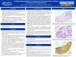
Case History: A 67-year-old male with a past medical history of alcohol abuse and previously treated Hepatitis C infection in 2012 with ledipasvir/sobosbuvir, was referred to our hospital for evaluation of hypercalcemia. Patient was in his usual state of health until 3 weeks ago when he started having fatigue, nausea, and anorexia. This was also associated with impaired memory and confusion. He was seen at an outside hospital and was found to have a calcium of 16.1 mg/dL (normal range 8.5-10.2 mg/dL), so he was given IV fluids, zoledronic acid and calcitonin with improvement in calcium to 10.8 mg/dL. Computerized tomography (CT) scan of chest/abdomen/pelvis showed a cirrhotic liver and a 6.5 cm hypodense mass within the left hepatic lobe. CT thoracolumbar spine was negative for lytic or blastic lesions with no acute fracture or dislocation. Patient was stabilized and transferred to our hospital for escalation of care. His parathyroid hormone (PTH) was low at 8 pg/ml (15-65 pg/ml) and PTHrP was high at 105 pg/ml (14-27 pg/ml). Repeat imaging with MRI showed a 17 x 8 cm area of signal abnormality with 2 more focal anomalies within, suspicious for malignancy which may be infiltrative. MRI thoracolumbar was negative for any metastasis, fractures or lytic lesions. Skeletal series was also negative for lytic lesions. Microscopic evaluation of the CT-guided liver biopsy showed two distinct patterns. A typical moderately differentiated HCC and a second malignant focus composed of hyperchromatic small to intermediate sized cells with apoptosis, atypical mitoses, vaguely palisading tumor cells around foci of necrosis. This second focus stained positively with CD56 and CAM 5.2 was suggestive of neuroendocrine differentiation and epithelial lineage. Background hepatic parenchyma showed early cirrhosis likely secondary to long standing hepatitis C or due to alcohol abuse. Tumor markers including alpha-fetoprotein (AFP), carcinoembryonic antigen (CEA), and carbohydrate antigen 19-9 (CA 19-9) were all negative. His workup was negative for another primary malignancy. His disease course was complicated by altered mental status and Klebsiella pneumoniae bacteremia. His PTHrP increased to 335 pg/ml. He received supportive care and expired 3 weeks from initial presentation.
Conclusion: Primary HCC and NEC generally tend to have a poorer prognosis than conventional HCC. To our knowledge, this case is the second report of primary mixed HCC and NEC associated with MAHC caused by the production of PTHrP.
Presentation Date
5-2020
Recommended Citation
Ishak, Rim S.; Entz, Abigail L.; Husain, Sanam; and Scher, Eric, "Combined Hepatocellular Carcinoma and Neuroendocrine Carcinoma Presenting with Hypercalcemia" (2020). Case Reports. 80.
https://scholarlycommons.henryford.com/merf2020caserpt/80
