

Files
Download Full Text (12.8 MB)
Program
Internal Medicine
Training Level
Resident PGY 2
Institution
Henry Ford Hospital
Abstract
Introduction: Sarcoidosis is a granulomatous T-cell-mediated disease with an incidence of 10.0 cases per 100,000 citizens per year in the U.S. Cutaneous involvement is seen in at least 33% of cases. The earliest case of tattoo-induced sarcoidosis was described in 1939. Now, predilection for tattoos is seen in primary Sarcoidosis and drug-induced sarcoidosis (DIS). DIS can occur in response to immune checkpoint inhibitors (ICIs), highly active antiretroviral therapy, Interferon therapy, Tumor Necrosis Factor-alpha antagonists and BRAF inhibitors. Nivolumab and Pembrolizumab are ICIs that inhibit programmed death protein 1 (PD-1), thus enhancing anti-tumor immunity, but also leaving patients susceptible to immune-related adverse events (irAEs) such as the development of autoimmune conditions.
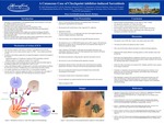
Case Presentation: A 78-year-old African American, non-smoking female with history of hypertension, hyperlipidemia and osteopenia presented with 2-month history of dry cough and 8 pound weight loss. She was not on ACE inhibitor for hypertension. CT chest showed a 4.0 x 3.1 cm right upper lobe mass with irregular borders and numerous enlarged intrathoracic lymph nodes. After tissue biopsy, she was diagnosed with stage IIIB lung adenocarcinoma. She harbored an exon 21 L858R EGFR mutation and was PD-1 negative. She had disease progression on separate Tarceva, Osimertinib and 6-cycle platinum-based regimens, so 4th-line Nivolumab was started. After 2 infusions of Nivolumab, 2 weeks apart, she developed a blue-green, non-erythematous induration at the site of her black-pigmented tattoo located on left lateral neck, inferior and posterior to left ear. She denied associated pain, scaling, bug bites, injections and trauma at the site. She received 3rd cycle of Nivolumab and the induration worsened. Dermatology obtained a biopsy of the tattoo lesion which showed non-caseating granulomas in the dermis, consistent with Sarcoid-type granulomatous dermatitis. She was not treated with steroids because her Indurated lesion resolved after discontinuation of Nivolumab.
Discussion: Diagnosing DIS remains difficult in oncology patients, as secondary causes must be ruled out. Solid organ and lymphoproliferative malignancies themselves can cause sarcoidosis but these cases involve B lymphocytes & histiocytes and are usually confined to the involved site, draining lymph nodes or metastatic sites5. Of the ICIs, DIS is least likely to be caused by PD-1 inhibitors and it is more common in melanoma malignancy4. This patient's onset after 2 weeks, is the earliest noted onset of Nivolumab-induced DIS ever reported. Previously, the earliest case reported was after 3 weeks. This is also the first tattoo-induced sarcoidosis in a patient with lung malignancy. Clinically, it is important to discern between DIS and progression of disease. In our patient, the onset of sarcoidosis after treatment, and resolution after discontinuing Nivolumab strongly suggests DIS. Hilar adenopathy in patients with suspected DIS should prompt bronchoscopy with biopsy and AFB smear to differentiate progression, sarcoidosis or other granuloma-causing conditions. Biopsy results can dictate changes in regimen versus continuation with current therapy.
Presentation Date
5-2019
Recommended Citation
Mohammed, Hadi and Mahmood, Sharmeen, "A Cutaneous Case of Checkpoint Inhibitor-Induced Sarcoidosis" (2019). Case Reports. 61.
https://scholarlycommons.henryford.com/merf2019caserpt/61
