

Files
Download Full Text (369 KB)
Program
Orthopedic Surgery
Training Level
Resident PGY 2
Institution
Henry Ford Hospital
Abstract
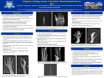
Purpose: Primary bone malignancies of the hand are rare as most cartilage lesions are small/ benign and can be managed with local curettage and grafting. Presented here is a small case series of malignant cartilage tumors of the hand. Because histologically their pathology overlaps and mimics their benign counterparts (enchondromas), it is of upmost importance to note the clinical presentation of potential malignant transformation. Case Series: Case 1: A 90 year old male who noted a slowly progressive, growing mass about his little finger proximal phalanx occurring in an area of a previously stable small mass. There was an associated decreased range of motion. Radiographs revealed an expansile mass with cortical disruption, fracturing and a periosteal reaction. MRI demonstrated a transcortical soft tissue mass. Patient was treated with a 5th ray amputation and pathology revealed a low grade chondrosarcoma. Case 2: A 54 year old woman presented with a progressively enlarging mass at her right ring finger distal phalanx that became occasionally painful after previously noting a lump for years. Radiographs revealed a lucency with calcification in the bone and MRI revealed periosteal reaction, cortical breakthrough and a soft tissue mass. The patient was treated with a mid-middle phalanx amputation and pathology revealed a low-to-intermediate grade chondrosarcoma with myxoid changes. Case 3: A 19 year old female with Ollier’s disease presented for routine follow up. Progressive dysfunction of the hand was noted after bumping the index finger of the left hand. Pain was improving but swelling was persistent. Physical exam revealed worsening hand function due to enlarging bulbous index finger. Due to dysfunction and growth index, ray amputation was performed. Pathology revealed a low grade chondrosarcoma with multiple other enchondromas noted. Discussion: Primary malignancies of the hand are rare whereas cartilage lesions, especially enchondromas, are common and rarely malignant. All patients in this series had preexisting small lesions with acutely progressive growth, and increasing but variable pain. Radiographs were consistent in showing cortical irregularity and were also suggestive of a soft tissue mass which was later confirmed on MRI. This is in contrast to our benign patients who typically have little or no pain and never exhibit a soft tissue mass. Conclusion: Awareness of the potential for chondrosarcomatous transformation is important to effectively manage this patient population. Notable soft tissue extension or enlarging masses, and pain in patients with preexisting cartilaginous neoplasms is needed to be considered as a sign of a biologically active and potentially malignant neoplasm.
Presentation Date
5-2019
Recommended Citation
Klag, Elizabeth; Easton, Matthew; Mott, Nicole; Buckley, Patrick; Evans, Timothy; and Mott, Michael, "Malignant Cartilage Lesions of the Hand: When Enchondromas Go Bad" (2019). Case Reports. 68.
https://scholarlycommons.henryford.com/merf2019caserpt/68
