

Files
Download Full Text (375 KB)
Program
Radiology - Diagnostic
Training Level
Resident PGY 3
Institution
Henry Ford Hospital
Abstract
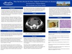
Background: Ovarian germ cell tumors (OGCT) represent between 1 in 5 and 1 in 4 ovarian neoplasms but are overwhelmingly benign with only a 5% malignancy rate. Within this category, mature teratomas (also known as ovarian dermoid cysts) are the most common type. And while they in fact represent the most common ovarian neoplasm in women age 30-40, they can occur at any age. The tumor is defined by its three germ cells layers: ectoderm, mesoderm and endoderm. This helps to characterize it on imaging with the presence of macroscopic fat on CT or MR considered diagnostic. Calcifications, including teeth, along with tufts of hair can also be seen. They are usually unilateral but are reported to be bilateral in 20% of cases and multiple can occur in a single ovary. Mature teratomas are generally slow growing and asymptomatic but can have mass effect and, if large enough, predispose to ovarian torsion. Another uncommon complication is rupture which can result in hemorrhage and shock in the short term with adhesions secondary to chemical peritonitis a long-term threat. Malignant transformation is exceedingly rare. Case Presentation: Our patient was a 58-year-old female without any reported significant past medical history given the caveat that she had not seen a physician in “years.” Of note, her mother and a sister had been diagnosed with breast and ovarian cancer respectively. She came to the emergency department with a three-month history of left lower quadrant abdominal pain, abdominal distention and a 20-pound unintentional weight loss over that time span. Relevant laboratory values for her demonstrated leukocytosis with a white blood cell count of 42 k/uL. She also had an acute kidney injury with a serum creatinine of 6.5 mg/dL. A CT of the abdomen and pelvis with contrast was obtained for further evaluation. This demonstrated a 10 x 11 x 9 cm mass arising from the left adnexa with a fat fluid level and intraluminal calcium as well as a dermoid plug.It abutted the adjacent bladder. There was air in the structure, raising concern for infection secondary to fistula formation. Both ovaries, along with the mass, were surgically removed and confirmed as a mature teratoma under histopathologic analysis. However, there was also invasion of the bladder and a sample from this invasive tissue demonstrated malignant transformation to squamous cell carcinoma. Discussion: Malignant transformation of mature teratomas are exceedingly rare occurring at a rate between 0.2-2.0 %. It becomes more common in older patients (mean age of patients with malignant transformation is 50 years old as compared to 33 years old with benign tumors). Tumors that are over 10 cm or that grow rapidly are also more likely to turn malignant.If the tumor is under 6 cm, management is usually conservative whereas bigger tumors are surgically excised with the goal of preserving normal ovarian tissue. The most common malignant cell type is indeed squamous cell cancer (SCC) as in our patient. Treatment is tailored to the particular transformative cell. This is an important case because it demonstrates an uncommon presentation of a common finding. As discussed above, mature teratomas are common ovarian entities and often have no significant clinical implications. But, as this case indicates, playing the odds is destined to fail eventually. Here, the size of the tumor and the weight loss were concerning clinical features. And on imaging there is not a definitive fat plane between the tumor and the bladder. The hoofbeats in this case looked like a horse on CT until put under the microscope.
Presentation Date
5-2019
Recommended Citation
Beckett, Ryan, "When The Horse Becomes the Zebra: Malignant Transformation of a Teratoma" (2019). Case Reports. 86.
https://scholarlycommons.henryford.com/merf2019caserpt/86
