

Files
Download Full Text (431 KB)
Program
Cardiovascular Disease
Training Level
Fellow
Institution
Henry Ford Hospital
Abstract
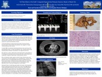
Introduction: It is extremely rare for leiomyosarcomas to affect the cardiovascular system. High degree of suspicion is required to diagnose this tumor in patients presenting with symptoms suggestive of a cardiac etiology. Because of the high mortality associated with this malignancy, early and aggressive intervention is crucial. Furthermore, imaging modalities may not adequately identify this tumor, as will be presented in this case leading challenges in diagnosis. Case: We present a case of a 59 year old female with a history of hypothyroidism who presented with progressive exertional dyspnea and palpitations. She underwent a chest CT which excluded pulmonary embolism but revealed diffuse long tubular narrowing above pulmonary valve involving main pulmonary artery raising suspicion forsupravalvularpulmonic stenosis. A 2D echocardiogram revealed normal left and right ventricular function, elevated systolic velocities distal to the pulmonic valve suggestive of supra-valvularpulmonary artery stenosis. A cardiac MRI was performed conforming pulmonary artery narrowing of the main pulmonary artery 1 cm above the pulmonic valve, with the narrowest area measuring 9mm in diameter. The pulmonic valve appeared uninvolved. She underwent a cardiac catheterization which demonstrated a peak gradient of 67 mm Hg across the stenotic lesion in the main PA. She was then diagnosed as symptomatic congenital isolated supra-valvularpulmonic stenosis. She was referred for cardiac surgery for relief of supra-valvularstenosis and reconstruction of the main pulmonary artery. Intraoperatively, a segment of the pulmonary artery was circumferentially narrowed by an infiltrative process. Frozen section analysis confirmed sarcoma, possible spindle cell variant. The main pulmonary artery was resected to the level of the pulmonary artery bifurcation, and a 23 mm aortic homograft was sewed in place. Subsequent biopsy revealed high grade spindle cell sarcoma, with morphologic features suggestingleiomyosarcoma. Re-review of the CT and MRI failed to conclusively predict the presence of this encircling tumor around pulmonary artery. Following surgery and recovery, she underwent a PET scan which demonstrated a small lytic lesion at L1, with possible metastatic femoral neck lesion. She was seen by hematology/oncology with recommendations to undergo localized radiotherapy and chemotherapy. Patient delayed treatment for her sarcoma in anticipation of a second opinion, and she ultimately passed away. Discussion: Leiomyosarcoma involving the pulmonary artery is extremely rare and usually manifests as a filling defect involving the pulmonary artery, mimicking a pulmonary embolism. In this case, multiple imaging studies were performed including a CT, MR and TTE, all of which failed torevealedthe extrinsic circumferential compression of the main pulmonary artery. In patients that do not have a congenital cardiac history (iepulmonic stenosis), a high degree of suspicion is required to rule out extrinsic compression by a tumor, as was evident in our case.
Presentation Date
5-2019
Recommended Citation
Saleh, Ashraf; Chamogeorgakis, Theistokles; Eng, Marvin; Yazigi, Firas; Stone, Chad; and Ananthasubramaniam, Karthik, "Congenital Supravalvular Pulmonic Stenosis, Maybe or Maybe Not" (2019). MERF 2019 - Case Reports. 3.
https://scholarlycommons.henryford.com/merf2019caserpt/3
