

Files
Download Full Text (576 KB)
Program
Radiology - Diagnostic
Training Level
Resident PGY 1
Institution
Henry Ford Hospital
Abstract
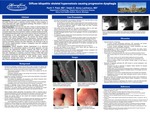
Background: There are a multitude of extrinsic and intrinsic etiologies of dysphagia, including mechanical obstruction, infections, neuromuscular conditions, and malignancies. Diffuse idiopathic skeletal hyperostosis (DISH) of the cervical and upper thoracic spine, a relatively common finding in spinal imaging, is rarely associated with upper esophageal pathology. We present a case of progressive dysphagia due to DISH. Case Presentation: A 64-year-old male presents to the emergency department with a three-day history of chest pain associated with cough, and vomiting. The patient admits to an extensive history of alcohol abuse with current everyday use and a 30-year smoking history. Upon further review, the patient noted increased weight loss and progressive inability to swallow liquids and solids associated with concurrent retrosternal chest pain and occasional vomiting. Initial cardiac workup was negative for any acute abnormalities. Follow up right upper quadrant ultrasound and HIDA scan were negative for acute cholecystitis. Dynamic swallow study showed silent aspiration of all consistencies. Follow up esophagogastroduodenoscopy was concerning for external compression of the esophagus at the upper esophageal sphincter. Subsequent imaging of the spine revealed bulky, flowing osteophytes in the cervical and thoracic spine consistent with diffuse idiopathic hyperostosis with varying degrees of airway and esophageal indentation throughout. At the time of publication, patient remained nil per os with nutrition supplementation through a nasogastric tube pending neurosurgical evaluation. Conclusions: Diffuse idiopathic skeletal hyperostosis, also known as Forestier’s disease, is an abnormal calcification of the anterolateral aspects of the spinal ligaments, and less commonly, the appendicular skeleton. The formation of bridging osteophytes through at least four consecutive vertebral bodies is required for diagnosis. Patients with DISH are usually asymptomatic, but can present with limited cervical mobility, or neck and back pain. Dysphagia is a relatively uncommon finding in DISH, but has been estimated to occur in up to 20% of cases. Mild cases are treated conservatively with physical therapy and pain control; however, progressively worsening symptoms or focal deficits require surgical management. Studies have shown recurrence of osteophytic lesions after surgical management is very common.
Presentation Date
5-2019
Recommended Citation
Patel, Parth Y. and Abreu Lanfranco, Odaliz E., "Diffuse idiopathic skeletal hyperostosis causing progressive dysphagia" (2019). MERF 2019 - Case Reports. 80.
https://scholarlycommons.henryford.com/merf2019caserpt/80
