

Files
Download Full Text (573 KB)
Program
Radiology - Diagnostic
Training Level
Resident PGY 3
Institution
Henry Ford Hospital
Abstract
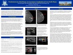
A 61-year-old female presented with persistent pain in the left breast following benign core needle biopsy. Diagnostic mammogram demonstrated a new small circumscribed mass in the upper outer quadrant of the left breast directly adjacent to the previously biopsied mass. Subsequent targeted Color Doppler ultrasound revealed a vascular mass with to and fro pulsatility. A diagnosis of breast pseudoaneurysm was made, a known albeit rare complication of breast core needle biopsy. The patient was treated with ultrasound guided thrombin injection into the pseudoaneurysm sac which resulted in complete sac thrombosis.
Presentation Date
5-2019
Recommended Citation
Davenport, Alexis A.; Williams, Paul; Pickney, David; and Segel, Mark, "Pseudoaneurysm of the Breast: An Uncommon Complication of Core Needle Biopsy" (2019). MERF 2019 - Case Reports. 85.
https://scholarlycommons.henryford.com/merf2019caserpt/85
