

Files
Download Full Text (516 KB)
Program
Orthopedic Surgery
Training Level
Resident PGY 4
Institution
Henry Ford Hospital
Abstract
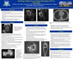
Introduction: Swelling and pain in a lower extremity is a common presenting complaint to the emergency room. Work-up often includes radiographs and ultrasound to rule out a deep vein thrombosis (DVT). Unfortunately, vascular ultrasounds which are negative for DVT may be mistaken as a definitive anatomic study by treating physicians. It is essential for physicians to consider that underlying malignancies, including soft tissue sarcomas, may be the cause of the patient’s symptoms – and that a vascular ultrasound cannot adequately assess for these lesions. Maintaining an appropriate differential diagnosis when performing subsequent evaluations is imperative. We present two unique cases of unilateral leg swelling which were initially evaluated by ultrasound and which were ultimately revealed to be caused by malignancy. Case Reports: Case 1: A 25-year-old pregnant female presented to the emergency room with left leg pain. Ultrasound at that time was negative for DVT, though a 3 x 4 cm hypervascular mass was noted in area. Lymph node exam upon follow-up was negative. The patient returned to the emergency room twice in the following months with repeat ultrasounds showing a progressively enlarging mass. Due to the patient’s pregnancy further imaging was deferred. Eventually, a CT scan revealed a solid mass, and orthopaedic surgery was consulted. Staging studies revealed disseminated metastatic synovial cell sarcoma. The patient was treated with radiation and chemotherapy and is currently alive with disease. Case 2: A 70-year-old male with history of renal cell carcinoma status post nephrectomy, prostate cancer, and bilateral arthritis of knees presented to his primary care doctor with unilateral calf swelling and pain. Vascular ultrasound was performed and was negative for DVT, with a hypoechoic mass noted measuring 3.1 x 1.6 cm. Vascular surgery evaluated the patient and no intervention was recommended. The patient had progressive swelling and new plantar surface numbness and was seen by orthopaedic surgery. An MRI revealed a 7.3 x 11.5 x 27.7 cm calf mass and a chest radiograph revealed multiple pulmonary metastases. Biopsy and staging work-up revealed high grade myxofibrosarcoma with disseminated metastatic disease. Patient is currently experiencing progressive disease.Discussion:Painful lower extremity swelling is a common chief complaint in the emergency room. Ultrasounds are used for evaluation of DVT and frequently the report is interpreted as only negative or positive for DVT. It is imperative to read the full report and to evaluate any potential reported masses with appropriate additional advanced imaging. Doing so can help to eliminate long lag times in diagnosing possible underlying soft tissue sarcomas. Conclusion: Emergency room vascular ultrasound of an extremity to rule out DVT is not satisfactory to rule-out an underlying malignancy. An appropriate degree of suspicion with directed anatomic imaging of the limb needs to be undertaken to avoid diagnosis delay of potential soft tissue sarcomas.
Presentation Date
5-2019
Recommended Citation
Evans, Timothy; Fisk, Felicity; Mott, Nicole; Buckley, Patrick; Easton, Matthew; and Mott, Michael, "Unilateral Leg Swelling with Negative Deep Vein Thrombosis by Ultrasound" (2019). MERF 2019 - Case Reports. 70.
https://scholarlycommons.henryford.com/merf2019caserpt/70
