

Files
Download Full Text (9.1 MB)
Program
Dermatology
Training Level
Resident PGY 4
Institution
Henry Ford Hospital
Abstract
History: A 5-day old black male full-term neonate born via vacuum-assisted delivery for non-reassuring fetal heart rate presented with congenital presentation of two asymptomatic midline lesions which appeared asymptomatic. There was no history of seizures, ophthalmologic findings, abnormalities in head circumference, height, weight or limb size. Newborn screening examination was unremarkable.
Examination: On the midline submental chin there was a soft, brown dome-shaped plaque measuring 0.8-centimeters with a circumferential ring of light brown pigmentation; on the midline upper chest there was a light brown 2-millimeter dome-shaped papule.
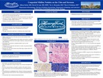
Course and Therapy: Ultrasound of the submental chin lesion revealed a 0.5 x 0.8 x 0.4-centimeter heterogeneously hypoechoic structure with a peripheral soft tissue rind. Punch biopsies of the submental chin and the midline upper chest revealed haphazardly arranged striated muscle fibers in the dermis, some of which inserted directly into the epidermis. The muscle fibers were highlighted by Masson’s trichrome and myogenin. Alcian blue revealed increased dermal mucin.
Discussion: Striated muscle hamartomas (SMH) are rare, benign congenital skin tumors characterized by haphazard arrangement of mature striated skeletal muscle, collagen, nerve bundles, and adipose tissue in the dermal and subcutaneous tissue. Although a rare entity, it is important to recognize this benign hamartoma as a congenital midline defect. Conservative management with clinical monitoring is recommended if cosmetically acceptable, as spontaneous regression over a period of years has been reported. Surgical excision may be pursued; however, the hamartoma may recur.
Presentation Date
5-2020
Recommended Citation
Zarbo, Allison; Luk, Kevin M.; Shwayder, Tor; and Friedman, Ben J., "Congenital Midline Nodules on the Chin and Sternum" (2020). Case Reports. 1.
https://scholarlycommons.henryford.com/merf2020caserpt/1
