

Files
Download Full Text (1.0 MB)
Program
Infectious Diseases
Training Level
Fellow
Institution
Henry Ford Hospital
Abstract
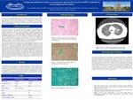
One week prior to demise, a 30 years old smoker male with a past medical history significant for intermittent asthma presented to emergency with shortness of breath, wheezing, productive cough, and generalized fatigue for 1 week. He was afebrile, normotensive, tachycardic and had O2 saturation of 96% on room air. Physical examination showed cachexia, audible wheezes and oropharyngeal erythema. Labs showed WBC 3600/uL with lymphocyte count of 700/uL and mild thrombocytopenia. Chest X ray was clear. Serology was reactive for HIV, pending viral load. Working diagnosis was asthma exacerbation in the setting of a possible viral infection for which he was discharged home to complete a 5-day course of high dose prednisone, with follow up with infectious diseases as an outpatient. Subsequent, HIV viral load after discharge was 194,643 copies/mL. One week later, he presented to the ED with worsening respiratory symptoms, new onset chest pain and vomiting. He was hypotensive, tachycardic, tachypneic, and afebrile. He had leukocytosis of 12,700/uL with neutrophilia, lactate of 6.6, BNP 841. Influenza A, B and RSV, and urine histoplasma antigen were negative. EKG showed abnormal ST segment elevation with concerns for STEMI. Chest CT revealed multifocal, bilateral ground glass and nodular opacities with cystic cavities. Mediastinal and hilar lymphadenopathy was also noted. Pulmonary embolism and pneumothorax were ruled out. Blood gases reflected acute hypoxemic respiratory failure. Vancomycin and Piperacillin/tazobactam were started. A bed side ultrasound showed significantly dilated right ventricle with severely reduced function and hence concerns for cardiogenic component of shock. He was intubated and shortly after developed asystole and expired after prolonged cardiopulmonary resuscitation within twelve hours of admission. At autopsy, gross exam showed bilateral pulmonary congestion, bilateral hilar adenopathy and matted lymph nodes in the mediastinum. Microscopy revealed cryptococcus (mucicarmine positive encapsulated yeast forms) involving intraalveolar and alveolar septal parts of all lobes of the lungs, effacing lymph nodes, and involving microscopic foci in bilateral myocardial ventricles. Modified GMS-positive cup shaped Pneumocystis organisms involved the alveoli of all lung lobes. The lung parenchyma showed minimal inflammatory response. Our case is of an HIV patient with respiratory symptoms found to have pulmonary co-infection with PJP and Cryptococcus neoformans, confirmed on pathology report. This is uncommon in literature. Additionally, this case is unique in reporting the presentation of Cryptococcus neoformans as involving the mediastinal lymph nodes and myocardium.
Presentation Date
5-2020
Recommended Citation
Tariq, Zain; Zia, Shereen; Schultz, Daniel; Brar, Indira; and Tibbetts, Robert J., "A unique presentation of Cryptococcus neoformans and Pneumocystis jirovecii PJP ) co infection in a newly diagnosed HIV patient" (2020). Case Reports. 12.
https://scholarlycommons.henryford.com/merf2020caserpt/12
