

Files
Download Full Text (552 KB)
Program
Emergency Medicine
Training Level
Resident PGY 1
Institution
Henry Ford Hospital
Abstract
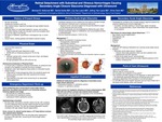
A 90-year-old female with past medical history of trigeminal neuralgia presented with a four-day history of a left-sided headache, nausea, vomiting, and vision loss in her left eye and one month of intermittent flashes of light in her left eye. Her left eye was diffusely injected with a cloudy cornea and fixed, mid-dilated, and non-reactive pupil. The vision in her, right eye was 20/200 with an intraocular pressure (IOP) of 16 mm Hg; her left eye was no light perception (NLP) with an IOP of 56 mm Hg. She was started on dorzolamide, brimonidine, and latanoprost eye drops. A bedside ultrasound performed by an emergency medicine physician demonstrated evidence of vitreous hemorrhage and concern for retinal detachment. Slit lamp examination performed by ophthalmologist demonstrated the left anterior chamber to be flat with a bulging iris and detached retina. Consequently, the patient was diagnosed with acute angle closure glaucoma secondary to increasing posterior chamber pressures. Given concern for altered mental status, the patient received a CT head in association with an inpatient MRI for headache refractory to home carbamazepine dosing regimen. Both imaging modalities corroborated the ultrasound's findings. In addition to the IOP-lowering medications, atropine, traditionally contraindicated in primary acute angle closure glaucoma, was added. Given her age, length of symptoms, and lack of light perception at presentation, her vision was deemed unsalvageable. Her pain was controlled with oral opioids and she was discharged with outpatient ophthalmology follow-up. At time of discharge, the IOP in her left eye was 49 mm Hg.
Presentation Date
5-2020
Recommended Citation
Holbrook, Michael B.; Kaitis, Daniel; Van Laere, Lily; Van Laere, Jeffrey; and Clark, Christopher R., "Retinal Detachment with Vitreous Hemorrhage Causing Acute Angle Closure Glaucoma" (2020). Case Reports. 127.
https://scholarlycommons.henryford.com/merf2020caserpt/127
