

Files
Download Full Text (427 KB)
Program
Family Medicine
Training Level
Resident PGY 2
Institution
Henry Ford Macomb
Abstract
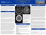
Tumefactive multiple sclerosis is a rare variant of multiple sclerosis that poses both diagnostic and therapeutic challenges for medical providers. This variant occurs infrequently, with an incidence of approximately 1-3 per 1000 cases of multiple sclerosis, with clinical features that make it difficult to distinguish from CNS neoplasm, infection, or infarction. Symptoms are highly variable, depending on areas of the brain affected, possibly impacting sensory, motor, and cognitive function. Serum labs may not reveal any specific abnormality, though CSF studies often contain oligoclonal band cells. Typical radiologic features identified in tumefactive MS may include the following; singular or multiple contrast enhancing lesions that are greater than 2 cm, incomplete to complete ring enhancement, lesion hypoattenuation on CT, hyperintense lesion on T2 MR, with minimal to no mass effect.In this case, a 39 year old caucasian male with no significant medical history, presented with acute onset right upper extremity weakness. Initial evaluation ruled out ischemic etiology or infectious etiology, however a comprehensive neurologic workup was delayed. The patient presented again for evaluation when his symptoms worsened and began to affect his right lower extremity. MR imaging was obtained, which demonstrated a 4.8 x 4.8 x 5.2 cm T2 hyperintense lesion within the left frontal lobe, that extended across the corpus callosum into the right frontal lobe. The characteristics of the lesion were concerning for malignancy and further workup was initiated. Subsequent CSF studies failed to demonstrate malignant cells, but did show oligoclonal band cells. Biopsy demonstrated numerous foamy macrophages with decreased myelin and preserved axons. Both of these findings being more consistent with a demyelinating process, than a malignant one, treatment with steroids was started for suspected tumefactive multiple sclerosis, and the patient symptomatically improved. Despite initial good response, the patient clinically declined and was started on disease modifying therapy (DMT), natalizumab. The patient again improved, but clinically declined, with repeat imaging concerning for expansion of the initial lesion. To help distinguish between tumefactive MS and malignancy, higher sensitivity Thallium MR was obtained, which was not suggestive of malignancy. The patient did subsequently improve after additional IV/PO steroid courses, as well as plasmapheresis, and has since been started on a second DMT (rituximab).This patient's presentation and course highlights the difficulty in distinguishing tumefactive multiple sclerosis from other CNS processes, as the initial symptoms and diagnostic features can have significant overlap. Response to steroids when clinical suspicion is high for tumefactive MS, can often help in differentiation of the underlying disease process and with symptoms. DMT is typically needed at some point in the patient's clinical course.
Presentation Date
5-2020
Recommended Citation
Macon, David, "Tumefactive Multiple Sclerosis, an Uncommon Variant with Several Mimics" (2020). Case Reports. 54.
https://scholarlycommons.henryford.com/merf2020caserpt/54
