

Files
Download Full Text (1.1 MB)
Program
WSU Medical School
Training Level
Medical Student
Institution
Wayne State University
Abstract
Introduction: Mucormycosis is a rare fungal infection most commonly seen in patients with neutropenia, diabetic ketoacidosis, or prolonged use of corticosteroids. Here we present an unusual case of isolated cerebral mucormycosis in a young, immunocompetent patient.
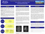
Case: A 36 year-old female with chronic Hepatitis C and intravenous drug abuse (IVDA) presented with sudden hemiparesis of the left upper and lower extremities. Her Glasgow Coma Scale was 15. MRI brain revealed a gadolinium-enhancing lesion adjacent to the right basal ganglia and thalamus consistent with an abscess, with surrounding vasogenic edema, mass effect causing right-to-left midline shift involving her right lateral ventricle (Figures 1 and 2). The abscess was surgically debrided and biopsy of the lesion revealed multinucleated giant cells and scattered groupings of branching fungal forms consistent with Rhizopus oryzae infection. Her remaining infectious and immunologic workup was unremarkable, including undetectable Hepatitis C RNA and absent HIV antibodies. During her hospital course, she exhibited new neurological findings including right lateral gaze palsy, left facial weakness, left hemi-neglect, and left ankle clonus. This was attributed to worsening vasogenic edema despite a medical regimen including IV amphotericin B and anidulafungin. Subsequently, isavuconazole was added. She continued on this regimen for four weeks after her initial debridement and was discharged to an in-patient rehabilitation center. On follow up examinations, she has exhibited gradual improvement in her strength, however she continues to have visual field deficits and requires assistance with activities of daily living.
Discussion: Mucormycosis is a rare, life-threatening fungal infection predominantly affecting immunocompromised hosts. The most significant risk factors for this infection include diabetic ketoacidosis, neutropenia, prolonged high-dose glucocorticoid therapy, bone marrow or solid organ transplantation, iron overload, and IVDA. Clinical presentation often reflects underlying risk factors for disease. For example, diabetic patients are likely to present with sinus involvement whereas patients with transplanted organs and malignancy more commonly present with pulmonary infection. Disseminated disease is most commonly seen in patients in an iron-overloaded state or those treated with deferoxamine. Isolated cerebral infection constitutes just 5% of all mucormyosis infections, but has a fatality rate exceeding 60%. Despite its rarity, however, isolated cerebral mucormycosis is the most common presentation among IVDA, constituting 62% of mucormycosis infections in this subgroup. Brain lesions are most commonly located within the basal ganglia, likely secondary to hematogenous seeding of the perforating branches of the Middle Cerebral Artery. It has been hypothesized that illicit injection drugs contaminated with mucormyosis spores enter the systemic arterial circulation before seeding the brain. Improved survival rates are seen among those patients treated with early stereotactic biopsy and aggressive amphotericin B therapy.
Conclusion: For patients with a history of IVDA who present with brain abscess (especially in the basal ganglia), mucormycosis should be on the clinicians’ differential diagnosis. Early intervention with stereotactic brain biopsy and amphotericin B, as well as abscess debridement, can dramatically increase the likelihood of survival and can minimize permanent neurologic sequelae.
Presentation Date
5-2020
Recommended Citation
Lewitt, Tessa and Rathnam, Anirudha S., "A case of Rhizopus infection in an immunocompetent IVDA host" (2020). Case Reports. 58.
https://scholarlycommons.henryford.com/merf2020caserpt/58
