

Files
Download Full Text (684 KB)
Program
Dermatology
Training Level
Resident PGY 2
Institution
Henry ford Hospital
Abstract
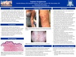
CASE REPORT: A 17-year-old healthy Latina female presented with an asymptomatic slowly enlarging birthmark. A purplish patch had been present over her left chest since birth and over time had gradually spread to involve her left shoulder and arm. She notes that sometimes the lesions will become darker red with paler colored skin at the periphery. There was no family history of similar findings.Over the left chest, shoulder, and ventral arm in a segmental distribution were numerous violaceous, vascular appearing macules coalescing into patches. The lesions were partially blanchable. Some lesions exhibited mild pallor at the periphery.A punch biopsy of the left arm displayed dilated and engorged thin-walled vessels in the papillary dermis, along with a mild perivascular lympho-histiocytic infiltrate with rare neutrophils.The initial differential diagnosis included angioma serpiginosum, unilateral nevoid telangiectasia, and acquired port wine stain. Given the history, distribution, clinical appearance, and pathology she was given a diagnosis of angioma serpiginosum. DISCUSSION: Angioma serpiginosum (AS) is a rare vascular nevoid disorder of unknown etiology. It has a female predominance (9:1) with onset during the first two decades of life. Most cases are sporadic, although an autosomal dominant inheritance pattern has been reported in two families. As is characterized by asymptomatic, pinpoint, violaceous to erythematous macules which may become papular. Slow progression in a serpiginous pattern can occur over time and partial involution may occur. Any area can be affected, but the buttocks, upper and lower extremities are most common. The distribution tends to be unilateral, sparing the palms, soles, and mucous membranes. AS must be differentiated from pigmented purpuric dermatosis (PPD), acquired port-wine stain (APWS), and unilateral nevoid telangiectasia (UNT). Histologically, AS is characterized by increased numbers of dilated capillaries in the upper dermis, which is identical to the pathology seen in UNT. In contrast to PPD, there is minimal inflammation, red cell extravasation, or hemosiderin deposition. APWS has a smaller degree of vessel proliferation than AS and usually follows a trigeminal nerve distribution. UNT is difficult to distinguish from AS both clinically and histopathologically. It is debated whether they represent distinct entities. Some authors report features favoring UNT include a characteristic dermatomal or Blaschkoid distribution (most commonly the C3-C4 and trigeminal regions) and anemic halos of vasoconstriction around individual telangiectasias. In contrast, the presence of lesions which are only partially blanchable favors AS. More definitive criteria may arise in the future as the field of medical genomics continues to expand. Management between UNT and AS is similar, consisting primarily of cosmetic treatment of the vascular ectasias with pulsed dye laser.
Presentation Date
5-2019
Recommended Citation
Robinson, Gabrielle; Schneider, Samantha; Veenstra, Jesse; Jahnke, Marla; and Douglass, Margaret, "A Teenage Girl with a Spreading Violaceous Birthmark" (2019). MERF 2019 - Case Reports. 15.
https://scholarlycommons.henryford.com/merf2019caserpt/15
