

Files
Download Full Text (602 KB)
Program
Ophthalmology
Training Level
Resident PGY 2
Institution
Henry Ford Hospital
Abstract
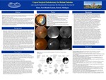
Purpose: Branch retinal artery occlusions can lead to devastating, permanent vision loss. Here we present a case of branch retinal artery occlusion (BRAO) treated with surgical embolectomy.Methods: A 67 year-old female presented with 14 hours of vision loss in the left eye. She had an ocular history of bilateral pseudophakia and bilateral pars plana vitrectomies (PPVs) for epiretinal membrane removal. Her medical history was notable for diabetes mellitus, hypertension, hyperlipidemia, and atrial fibrillation.Her visual acuity was 20/100 in the left eye. Confrontation fields showed a superior altitudinal defect in the left eye. Indirect ophthalmoscopy revealed a retinal embolus on the nasal aspect of the optic disc with inferior retinal whitening. Surgical embolectomy was performed 17 hours after symptom onset.After discussion of the potential risks and benefits of surgical embolectomy, the patient wished to proceed. Three hours after presentation, she was brought to the operating room, placed under general anesthesia, prepped and draped in the usual ophthalmic fashion. A surgical embolectomy was performed. Transscleral trocars were placed in the inferotemporal, superotemporal, and superonasal quadrants. The conjunctiva was displaced with 0.12 mm forceps, and sclerotomy sites were created 3.5 mm posterior to the limbus. Core vitrectomy was performed. Magnification was provided by a Hassan-Tornambe contact lens. Crushing of the embolus was attempted with a soft tip flute needle, but this was not successful. An arteriotomy with a 23-gauge microvitreoretinal (MVR) blade was attempted, but minimal hemorrhage occurred. Internal limiting membrane (ILM) forceps were used to attempt to crush the embolus, but this was also not successful. Next, curved scissors were used to make an arteriotomy, and a small hemorrhage appeared. ILM forceps were used to remove the embolus. Sclerotomy trocars were removed. Sclerotomy sites were ensured that they were water tight.At 8 weeks after surgery, the patient’s vision returned to near baseline (20/25). Fluorescein angiography showed near normal fluorescein transit time. Postoperative optical coherence tomography at one month showed resolved retinal edema, but nasal outer retinal and retinal pigmented epithelium atrophy. On postoperative one and two month visits, her 30-2 HVFs showed near complete superior altitudinal defects.Results: The retinal embolus was successfully removed. Her visual acuity returned to near baseline. Fluorescein angiography confirmed reperfusion. She retained a significant superior altitudinal defect.Conclusion: This case demonstrates the potential of embolectomy/embolysis to restore anatomical retinal perfusion in patients with fovea involving BRAOs with retinal emboli present on the optic disc. Large, multicentered prospective studies are needed to evaluate the efficacy and safety of surgical embolectomy for the acute management of fovea involving BRAOs.
Presentation Date
5-2019
Recommended Citation
Aung, Andre and Brill, Daniel, "Urgent Surgical Embolectomy for Retinal Embolus" (2019). MERF 2019 - Case Reports. 54.
https://scholarlycommons.henryford.com/merf2019caserpt/54
