

Files
Download Full Text (698 KB)
Program
Internal Medicine
Training Level
Resident PGY 1
Institution
Henry Ford Macomb
Abstract
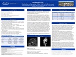
Introduction: Histiocytoses are a group heterogeneous diseases of unknown cause affecting myeloid progenitor cells. Erdheim-Chester disease (ECD) is a subclassification of Non-Langerhan cell Histiocytosis (LCH). ECD has characteristic lesions of the skeletal, cardiac, and vascular systems. There are many instances when LCH and ECD occurs concurrently, called mixed Histiocytosis. Fewer than 500 cases of ECD have been reported and even fewer of mixed histiocytosis. Case: 42 year old caucasian female presented after a syncopal event, status post tenosynovitis release surgery. Patient presented with pallor and hypotension upon arrival, prompting a syncope workup. Patient admitted long standing history of claudication, fever, and weight loss.Labs yielded a WBC of 10.5 ESR of 67, and CRP of 2.3. Initial CTA to rule out PE revealed inflammation of the aorta and left subclavian vessel with mural thickening of descending aortic arc. CTA also showed stenosis of the celiac, superior mesenteric, and renal arteries, suspicious of Takayasu arteritis. Subsequent CT and MRI exhibited a suprasellar mass which was later resected and biopsied diagnosed as BRAF positive Langerhans Cell Histiocytosis, staining positive for CD1a, S-100, Langerin, CD68 and CD168. Discussion: The BRAFv600e gene mutation is implicated in both LCH and ECD, suggesting a common origin. After discovery of the BRAF mutation on both LCH and ECD, a study reported significant co-occurrence between the two, including 19% of the largest ECD cohort. Recognizing LCH, ECD, and mixed histiocytosis is imperative as treatment regiments regimens differ. Thus, actively considering mixed histiocytoses is important in the setting of LCH diagnosis.
Presentation Date
5-2019
Recommended Citation
Papukhyan, Hayk; Kerndt, Connor; Gakhal, Gurinder; and Patel, Prashant, "Mixed Histocytosis Manifesting as Suprasellar Mass with Aortic Involvement" (2019). MERF 2019 - Case Reports. 97.
https://scholarlycommons.henryford.com/merf2019caserpt/97
