

Files
Download Full Text (484 KB)
Program
Diagnostic Radiology
Training Level
Resident PGY 2
Institution
Henry Ford Hospital
Abstract
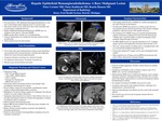
Hepatic Epithelioid Hemangioendothelioma (HEHE) is a rare, low to intermediate grade malignant lesion of vascular origin which has an incidence of less than 0.1 cases per 100,000 people. Given its rarity, this lesion is often misdiagnosed. We present a case of a patient diagnosed with HEHE who was initially thought to have NASH cirrhosis and hepatocellular carcinoma. Our case demonstrates the characteristic imaging features of this lesion and a discussion about the disease treatment and prognosis. The patient in our case was a 34 y/o male with a past medical history of DVT who presented with increased shortness of breath and weight gain which had been worsening over a period of months. He had no significant family history or history of alcohol use. Physical exam showed obesity and ascites. Laboratory testing showed negative HCV Ab, HBsAg, ANA, AMA, and AMSA. LFTs and AFP were normal. Ultrasound was ordered and showed steatosis and multiple small incompletely characterized liver masses. Given workup, concern for NASH related cirrhosis with possible HCC. Multiphasic CT and liver biopsy were negative for malignancy but showed evidence of venous outflow obstruction. Angiogram demonstrated severe stenosis of the IVC at the right atrial junction which was treated with angioplasty resulting in temporary relief in symptoms. However, the patient returned with increasing ascites. Repeat MRI showed multiple ring enhancing masses thought to be metastases or abscesses. Biopsy showed vascular neoplasm consistent with epithelioid hemangioendothelioma. Patient was then referred for orthotopic liver transplant. The imaging manifestations of HEHE are variable and depend on the stage of disease at presentation. If found early, HEHE may present as a solitary lesion, however most patients present with advanced disease and will have multiple peripheral, coalescent lesions with capsular retraction. On unenhanced CT, HEHE lesions are typically hypoattenuating without calcifications. Enhanced CT may show the characteristic “lollipop sign” caused by hepatic or portal vein branches which terminate within the edges of lesions. On MRI, HEHE lesions are typically hypointense on T1-weighted images and have heterogeneously increased signal intensity on T2. The most common pattern of enhancement is a peripheral halo with an occasional thin peripheral hypointense rim. Ferumoxide-enhanced T2 images can be used to distinguish tumor margins. Many reported cases of biopsy proven HEHE do not present with many of these characteristic imaging findings. Given its variable appearance, 60-80% of these lesions are initially misdiagnosed. Underlying comorbidities such as cirrhosis, found in only 1% of HEHE cases, can also confound diagnosis. Differentiating HEHE from its differential diagnoses such as hepatic metastatic carcinoma, cholangiocarcinoma, and other liver vascular tumors like hepatic angiosarcoma or cavernous hemangioma requires histopathological staining. No standard of treatment for HEHE exists given its rarity. As patients often present with multifocal disease, orthotopic liver transplantation is often performed. Adjuvant chemotherapy and radiation are also a part of the treatment regimen. Prognosis depends on whether or not extrahepatic involvement is present at the time of diagnosis with hepatic confined disease having a 1 and 5 year survival rate of 80% and 64% respectively.
Presentation Date
5-2020
Recommended Citation
Cormier, Peter; Kaddurah, Omar; and Hanson, Ronela, "Hepatic Epithelioid Hemangioendothelioma: A Rare Malignant Lesion" (2020). MERF 2020 - Case Reports. 105.
https://scholarlycommons.henryford.com/merf2020caserpt/105
