

Files
Download Full Text (434 KB)
Program
Pathology
Training Level
Resident PGY 4
Institution
Henry Ford Hospital
Abstract
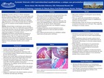
Systemic sclerosis is a chronic autoimmune disease of unknown etiology. Disease course usually begins with Raynaud’s phenomenon followed by skin sclerosis and internal organ involvement. Diagnosis is made based on the clinical symptoms, presence of antibodies and endoscopy with biopsy. With this background, we present a case of a 60-year female who was followed by GI clinic for treatment resistant dyspepsia, bloating and nausea. Past medical history was significant for fibromyalgia, Raynaud’s disease and transient ischemic attack. For evaluation of dyspepsia, she underwent endoscopy with gastric and esophageal biopsies, which were superficial and showed only non-specific chronic inflammation. Further, 24 hour pH monitoring results were unhelpful in making a definitive diagnosis. Esophageal manometry showed a hypertensive lower esophageal sphincter for which she underwent Heller’s myotomy. Her symptoms, however, persisted and she was scheduled to undergo laparoscopy and open gastric wall biopsy. Stomach biopsy revealed gastric mucosa showing focal vascular ectasia. There was significantly increased fibrosis involving the muscularis mucosae and propria (Figure A), highlighted by trichrome stain (Figure B-D). Following biopsy results, Scl-70 Ab test was performed and it turned out to be positive (42, Nunits). Thus, supporting the diagnosis of systemic sclerosis. Full thickness gastrointestinal tract biopsies of systemic sclerosis cases are rarely seen in routine surgical pathology practice. This case is unique because of the complexity of clinical presentation requiring open gastric wall biopsy but demonstrates the value of pathologic evaluation for diagnosis of rare autoimmune disorders such as systemic sclerosis.
Presentation Date
5-2020
Recommended Citation
Ahsan, Beena U.; Mehrotra, Harshita; and Ormsby, Adrian, "Systemic Sclerosis with Gastrointestinal manifestations: a unique presentation" (2020). MERF 2020 - Case Reports. 121.
https://scholarlycommons.henryford.com/merf2020caserpt/121
