

Files
Download Full Text (215 KB)
Program
Ophthalmology
Training Level
Resident PGY 3
Institution
Henry Ford Hospital
Abstract
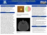
Traumatic optic neuropathy is a potentially visual devastating event caused by acute injury to the optic nerve from direct or indirect trauma. It usually presents with decreased vision and a relative afferent pupillary defect. Oddly, the dilated fundus exam is usually unremarkable. Treatment at this time is controversial with options including observation, corticosteroids, or surgery. A 47 year old male presented with decreased vision of left eye after falling down the stairs. Exam showed a vision of no light perception, 4+ relative afferent pupillary defect, and a normal fundus exam. CT showed displaced acute fracture of the medial wall and roof of the left orbit as well as a fracture of the left optic canal concerning for optic nerve compromise. Given patient had no contradictions, patient was started on IV solumedrol with a 30 mg/kg loading dose, followed by 5.4 mg/kg q 6 hours for 48 hours. Steroids are thought to reduce edema to help reduce optic nerve damage. Roughly 2 week later, the vision of the left eye improved to a vision of count fingers. This case report shows corticosteroids may play a role in helping to improve vision in the cases of traumatic optic neuropathy if no contradictions to steroids are present.
Presentation Date
5-2020
Recommended Citation
Gandhi, Sachin; Gandhi, Shawn; Brill, Daniel A.; and Yousif, Candice, "Corticosteroid Use in Traumatic Optic Neuropathy" (2020). MERF 2020 - Case Reports. 93.
https://scholarlycommons.henryford.com/merf2020caserpt/93
