

Files
Download Full Text (790 KB)
Department
Surgery; Thoracic Surgery
Position/Job Title
Resident; Physician
Description
Introduction: Bronchoscopy with associated mediastinal lymph node sampling and biopsy of pulmonary lesions is critical in the diagnosis of pulmonary disease, such as cancer or inflammatory conditions, as well as in oncologic staging. Biopsies via traditional flexible bronchoscopy have been limited to lesions easily accessible by the mainstem bronchi or larger segmental branches. Endobronchial ultrasound with fine needle aspiration (EBUS-FNA) of mediastinal lymph nodes or extraluminal lesions in central locations may also be performed. However, peripheral pulmonary lesions are often unable to be accessed easily or safely through flexible bronchoscopy. Computed Tomography (CT) guided transthoracic needle aspiration is an alternative method of biopsy of peripheral pulmonary lesions, but is also associated with a high rate of pneumothorax. The development of robotic bronchoscopy platforms, like the MonarchTM Platform and the IonTM endoluminal robotic bronchoscopy platform, has provided physicians access to peripheral pulmonary nodules in a way that was not previously provided by conventional bronchoscopy or CT-guided methods. Additionally, the diagnostic yield of bronchoscopy has increased to 80-90% using robotic technology. Today, however, there is a relative paucity of literature reporting the incidence and type of complications associated with this new technology. Studies to date have reported minimal adverse events, which have been limited to pneumothorax, bronchopulmonary bleeding, and acute respiratory failure, suggesting robotic bronchoscopy can be a safe and useful diagnostic procedure.
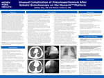
Case Report: This case report serves to detail a rare complication of robotic bronchoscopy, pneumoperitoneum. The patient was a 60 year old male who presented for diagnostic workup of a pulmonary nodule, suspected to be malignant, as well as placement of a percutaneous endoscopic gastrostomy (PEG) tube for malnutrition. Robotic bronchoscopy was utilized to perform needle aspiration of multiple peripheral pulmonary lesions, one of which abutted the diaphragm. Following bronchoscopy, abdominal distension was noted precluding safe PEG tube placement. Abdominal x-ray revealed large volume pneumoperitoneum. The patient was transferred to the surgical intensive care unit (SICU) and closely monitored. Ultimately, no operative intervention was indicated, and the patient was discharged to home after a period of observation.
Conclusion: Although a rare complication, it is critical for physicians to be aware of the possibility of pneumoperitoneum following peripheral biopsy via robotic bronchoscopy, as well as to be able to readily identify this finding and manage accordingly.
Publication Date
4-16-2024
Recommended Citation
Day, Ashley and Kulkarni, Mohan G., "Unusual Complication of Pneumoperitoneum After Robotic Bronchoscopy on the MonarchTM Platform" (2024). 2024 Henry Ford Jackson Hospital Research Symposium. 10.
https://scholarlycommons.henryford.com/hfjhrs2024/10
