

Files
Download Full Text (499 KB)
Department
Surgery; Center for General Surgery
Position/Job Title
Resident; Physician
Description
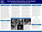
Hemorrhagic cholecystitis is a rare, and sometimes under considered, cause of acute cholecystitis. Hemorrhagic cholecystitis can be precipitated secondary to blunt abdominal trauma, malignancy, or various etiologies of bleeding or clotting disorder, including anticoagulation use, kidney failure, or liver failure. A combination of various imaging techniques and a high level of clinical suspicion is required to accurately diagnose hemorrhagic cholecystitis and prevent high-risk and possible complications that can result from delay in diagnosis and treatment. We present a case report involving spontaneous hemorrhagic cholecystitis, along with a review of imaging findings in various modalities. Our patient was a 69 y/o male with 12 hours of epigastric pain, nausea, and vomiting. Medical history was significant for venous thromboembolism currently on Coumadin. Lab results showed leukocytosis, elevated liver enzymes and bilirubin, and an INR of 4.1. Imaging with RUQ ultrasound and CT abdomen was consistent with acute cholecystitis with additional findings of internal debris, which raised suspicion for hemorrhagic cholecystitis. Management included reversal of anticoagulation followed by urgent minimally invasive cholecystectomy.
Publication Date
4-16-2024
Recommended Citation
Arscott, Tyler and Narkiewicz, Lawrence, "Hemorrhagic Cholecystitis: A Case Report" (2024). 2024 Henry Ford Jackson Hospital Research Symposium. 12.
https://scholarlycommons.henryford.com/hfjhrs2024/12
